LIBR MRI Neuroimaging Facility
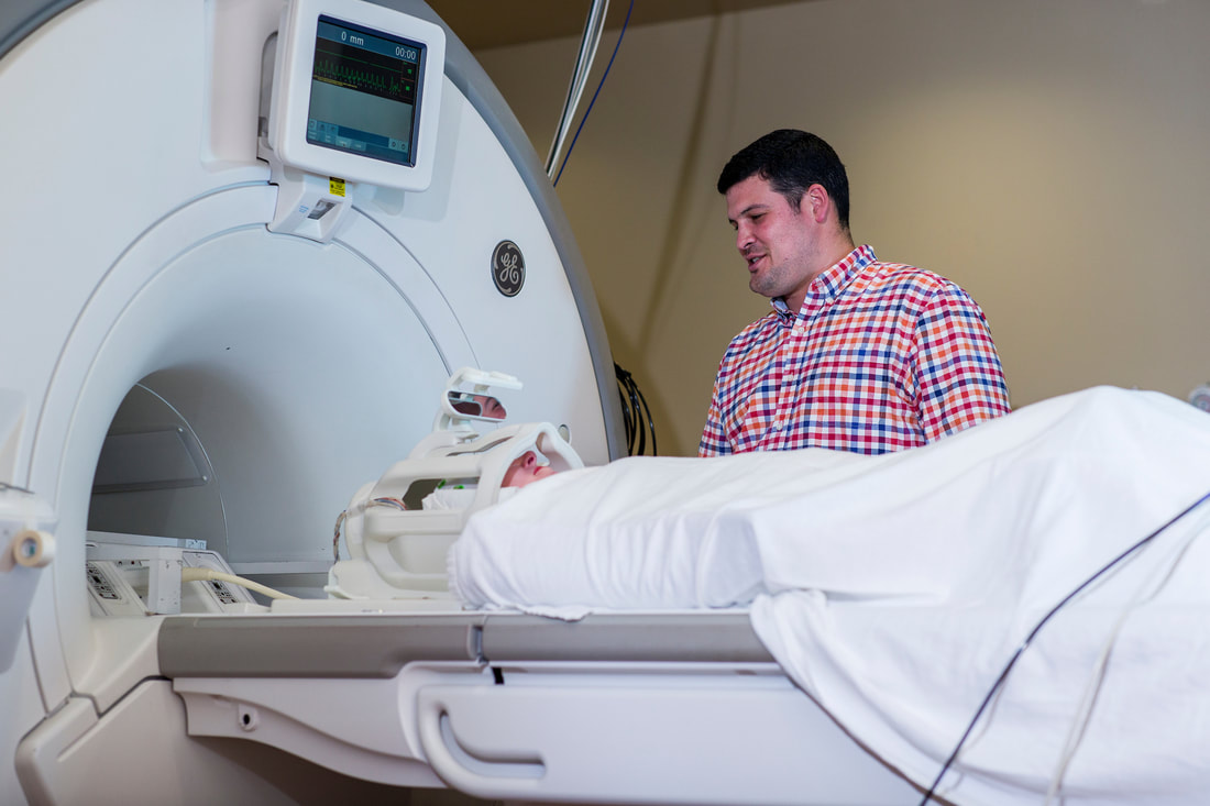
Established in July 2009 and in research operation since June 2010, the MRI facility provides advanced state-of-the-art MRI, functional MRI (fMRI), simultaneous electroencephalography (EEG) recording with fMRI, and real-time neuroimaging capabilities. Two MRI scanners are fully dedicated to research and provide an advanced capacity for the latest quantitative imaging of the human brain structure and online monitoring of brain activity in real-time. The two scanners can also be synchronized and integrated for hyperscanning, where EEG and fMRI signals of interacting two subjects are measured simultaneously.
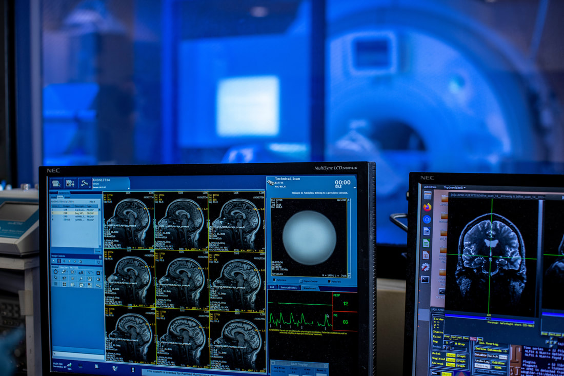
The facility provides all the latest technology, tools, and resources necessary to conduct and support brain neuroimaging studies focused on advancing clinical research to discover causes of and cures and novel interventions for mood, anxiety, eating, and memory disorders. In 2023 LIBR purchased a new state-of-the-art MR system, a Siemens 3.0 Tesla Prisma NX, to replace the original system installed in 2009. The arrival of this new system with state-of-the-art coils and methods will support more powerful imaging results for LIBR research in the coming decade. The new system is the focus of several NIH grants and LIBR projects awarded in 2023. It complements the ongoing work supported by the second scanner, a General Electric 3.0T Discovery 750. LIBR’s custom-made data management system allows for automatic handling of large amounts of neuroimaging data and real-time integration of fMRI, physiological data (respiration, pulse oximetry, or ECG waveforms), and EEG data simultaneously acquired with fMRI. These capabilities enable live monitoring of ongoing brain activation in real-time with extensive multimodal information. |
LIBR employs a custom real-time imaging system that provides continuous scan quality monitoring, resulting in superior functional MRI. This real-time system is also an integral part of a neurofeedback system, in which subjects are trained to self-modulate their thoughts and brain activity using feedback that is calculated from their own scans in real-time. Neurofeedback is being investigated as a potential treatment for psychiatric disorders. Neurofeedback also provides online optimization of brain stimulation with simultaneous transcranial direct or alternating current stimulation (tDCS/tACS) during fMRI.
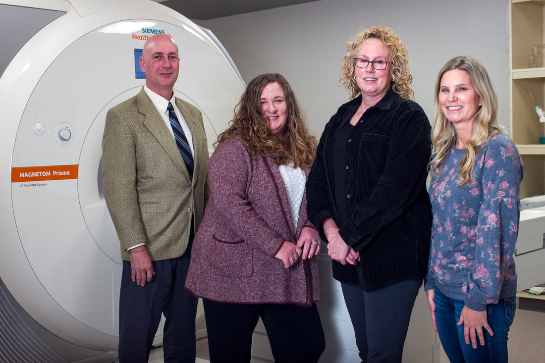
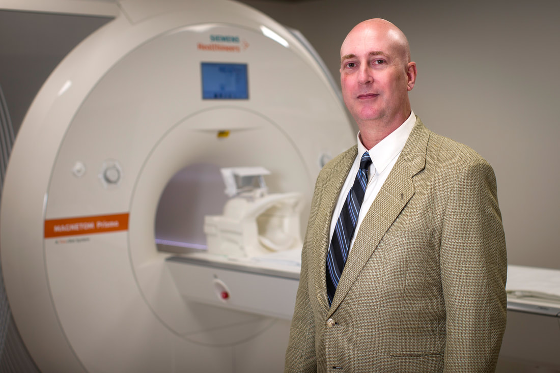
The advanced combination and customization of state-of-the-art MRI, RF coils, EEG, and brain stimulation technologies, along with custom-developed software solutions and a wide range of auxiliary computerized equipment, offer a unique potential for conducting advanced brain research. The LIBR MRI facility also supports collaborative neuroimaging research with researchers from local academic institutes, including The University of Oklahoma, Oklahoma State University, and The University of Tulsa. The MRI facility is directed by Michael Rohan, Ph.D., an expert in functional imaging systems and methods. It operates with the technical support of Masaya Misaki, Ph.D., an expert in multimodal neuroimaging data analysis and information technology and under the management of Julie Arterbury, Chief of MRI Technologist. Additional staff includes two MRI technologists, Leslie Walker and Amy Ginn. The MRI facility was founded and built by Jerzy Bodurka, Ph.D., and is named in his honor. |
|
|